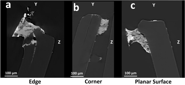
Figure 7.
XRM virtual cross-sections of growth sites. The Scout & Zoom protocol coupled with the state-of-art DeepScout DL reconstruction algorithm was employed to identify smaller VOIs that correspond to the edge, corner, and planar surface fungal growth sites. These selected VOIs were rendered at higher resolution (pixel size 0.75 μm). After examining the 2D slices in the YZ plane, which depict internal cross-sections of PET fragments, it becomes evident that F2 F. oxysporum was able to penetrate deeply into PET near an edge (a), or, somewhat less extensively, near a corner (b). On the other hand, on the flat surface, there was only superficial growth (c).

