Figure 2.
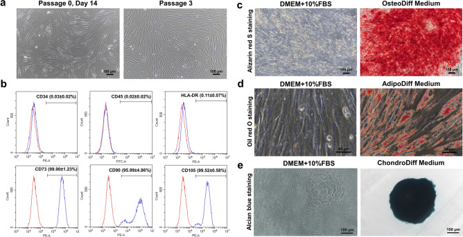
Characterization of human placenta-derived mesenchymal stem cells (PL-MSCs). (a) The spindle shape morphology of PL-MSCs cultured in DMEM supplemented with 10% fetal bovine serum on day 7 after removal of non-adherent cells (left) and in passages 3 (right). (b) Flow cytometric analysis of surface marker expression in PL-MSCs showing positive expression of MSC markers (CD73, CD90, CD105) and negative expression of hematopoietic markers (CD34, CD45, HLA-DR). (c) Brilliant orange-red staining of alizarin red S in PL-MSCs on day 28 of their osteogenic differentiation. (d) Positive signal of Oil Red O staining in PL-MSCs on day 28 on their adipogenic differentiation. (e) Chondrogenic differentiation potential of PL-MSCs demonstrated by Alcian positive blue color staining of positive colonies (right) that develop in the presence of chondrogenic differentiation media. Differentiated colonies were obtained from cells of all 5 donor placentas. (a) and (c) were captured with 10X magnification. Scale bar = 100 μm. (d) was captured with 40X magnification. Scale bar = 50 μm. (e) was captured with 20X magnification. Scale bar = 100 μm.

