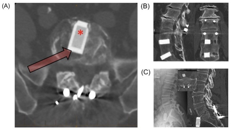
Figure 2.

Thirty-year follow-up after interbody fusion using silicon nitride ceramic spacers.10 (A) Computed tomographic (CT) image showing silicon nitride implant with load bearing nonporous rim (arrow) and porous core conducive to early osseointegration (asterisk). (B) Sagittal CT image showing fusion mass and osteointegration. The coronal CT image shows fusion mass adjacent to the spacer and osseointegration at the bone-ceramic conjunction. (C) Left: Implant anterior translation with posterior revision. Insert: L5 to S1 demonstrating fusion, despite anterior movement of implant. Right: Lack of reaction in surrounding tissue reinforces biocompatibility of silicon nitride implant. This image is reprinted with permission from Mobbs et al.14 Copyright 2018 Elsevier Publishing Group.
