FIGURE 1.

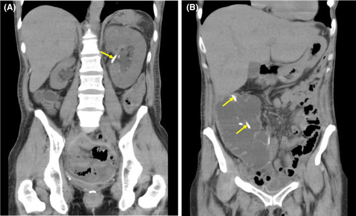
Plain computed tomography on the initial consultation. Bilateral mild hydronephrosis, right renal atrophy (A), and marked dilation of a urinary pouch (B) are observed. A ureteral stent (arrows) had been placed.

Plain computed tomography on the initial consultation. Bilateral mild hydronephrosis, right renal atrophy (A), and marked dilation of a urinary pouch (B) are observed. A ureteral stent (arrows) had been placed.