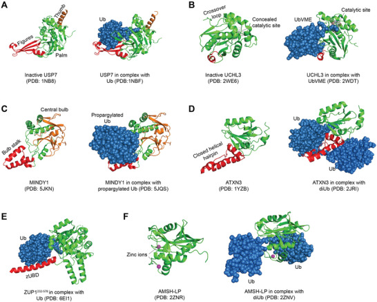
Figure 3.

Representative crystal structures of DUBs in cartoon. The crystal structures of USP7 (A), UCHL3 (B), MINDY1 (C), ATXN3 (D), ZUP1231–576 (E), AMSH‐LP (F) in the absence (left panel) or presence (right panel) of Ub substrates/inhibitors were adapted from protein data bank (PDB) using PyMOL software.
