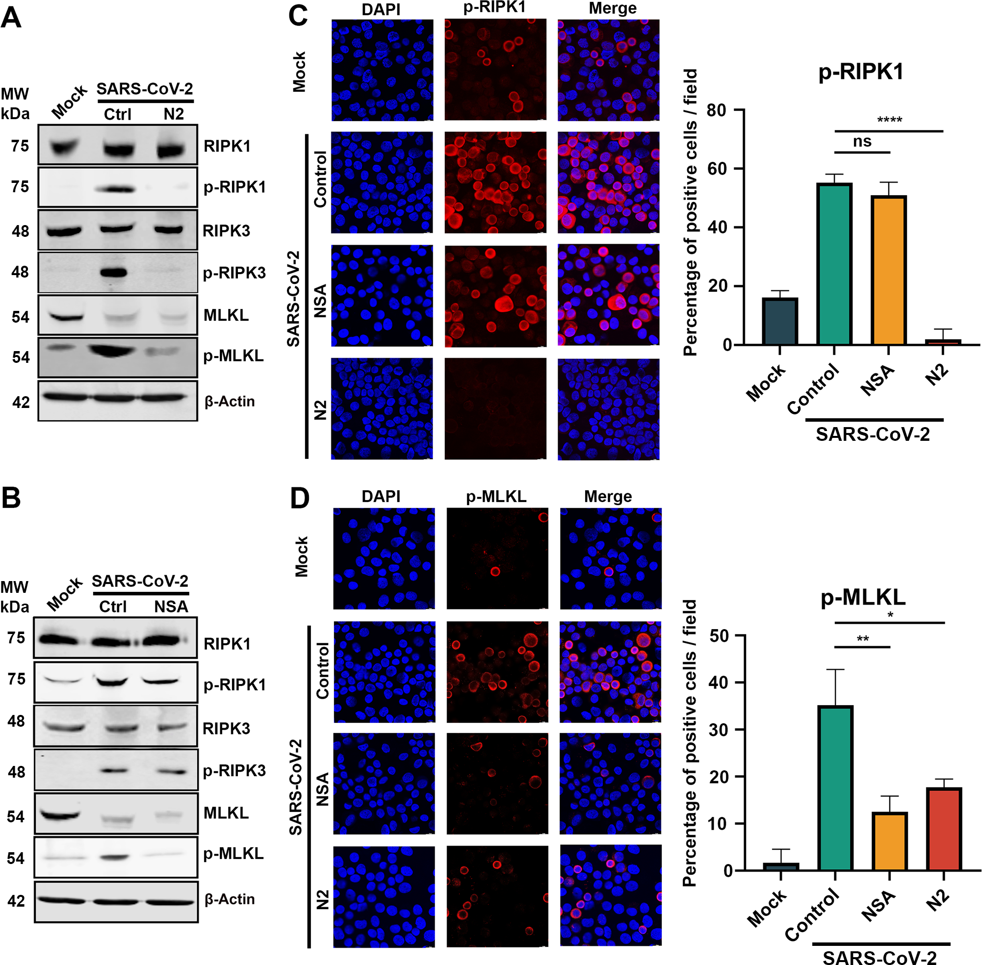
Figure 5. Both NSA and N2 inhibit necroptosis via RIPK1 to MLKL pathway.

CuFi-ALI cultures were treated with NSA (A) and N2 (B) at 20 and 50 μM, respectively, followed by infection of SARS-CoV-2 at an MOI of 2. DMSO was used as a vehicle Control (Ctrl). At 3 dpi, cell lysates were collected. (A&B) Western blotting. The lysates were separated on SDS-10%PAGE and blotted for the expression of both phosphorylated (p-) and unphosphorylated RIPK1, RIPK3, and MLKL. β-actin was detected as a loading control. (C&D) Immunofluorescence analysis. The collected cells were cytospun onto slides, stained with anti-p-RIPK1 (B) and anti-p-MLKL(D), respectively. Confocal images were taken at a magnification of × 63. Nuclei were stained with DAPI (blue). Positive stained cells were counted, and percentages of the positive cells were plotted at the right side of panels C&D.
