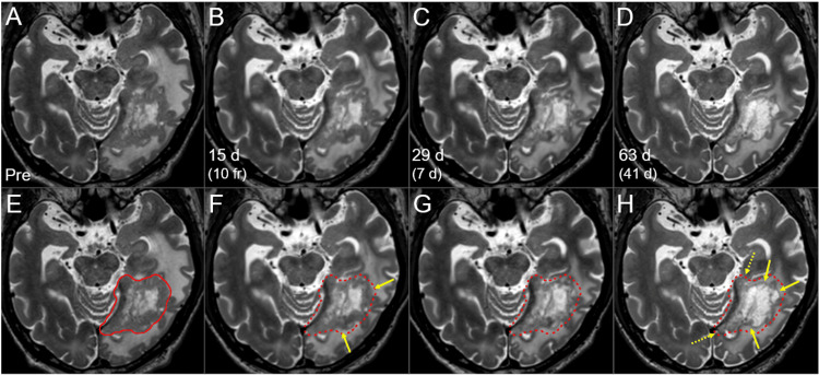
Figure 4. T2-WIs during the 15-fr radiosurgery and subsequent WBRT.
The images show (A-H) axial T2-WIs; (E-H) the GTV contour before the SRS (solid line in E, dashed lines in F-H) superimposed onto T2-WIs; (A, E) three days before the initiation of SRS; (B, F) 15 days after the SRS initiation (at 10 fr); (C, G) 29 days after the SRS initiation (seven days after the completion of SRS) and (D, H) 63 days after the SRS initiation (41 days after the SRS completion, the next day after the WBRT completion).
(A-H) These images are shown at the same magnification and coordinates under co-registration and fusion based on the pre-SRS images. The solid component of the lesion gradually decreased (arrows in F, H), except for the components near the tentorial incisura (dashed arrows in H).
d: days; WI: weighted image; GTV: gross tumor volume; SRS: stereotactic radiosurgery; fr: fraction(s); WBRT: whole-brain radiotherapy

