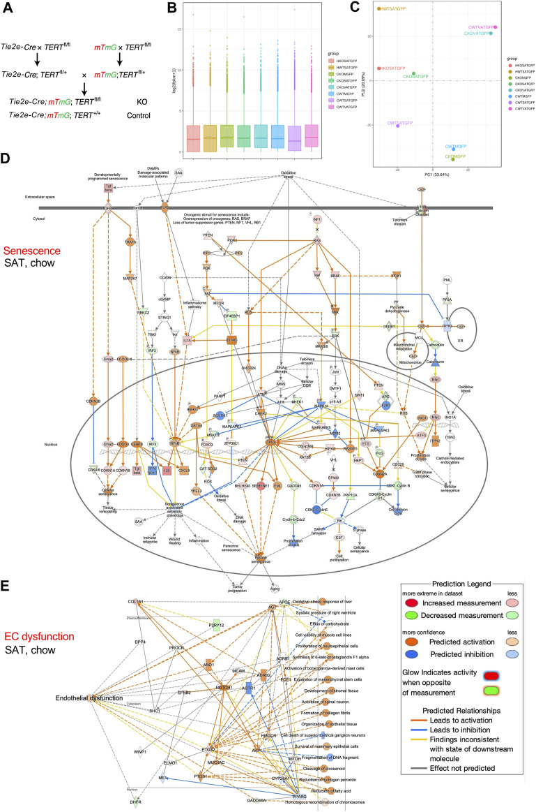
FIGURE 1.
TERT knockout in mouse endothelial cells (EC). (A), Breeding scheme to generate mice with mG+ and TERT-EC-KO (fl/fl) or WT (+/+) Tie2+ cells (EC) and other cells mT+. EC senescence and dysfunction caused by TERT loss was assessed in 8-month-old female mice fed chow (C) or HCD (H). Cells isolated from subcutaneous AT (SAT), intraperitoneal visceral AT (VAT), quadricep and gastrocnemius skeletal muscle (M) were subjected to FACS sorting to isolate mG+ cells for mRNA extraction. (B), Gene expression distribution. X axis: mouse groups (also shown on the right). Parameters of box plots are indicated, including maximum, upper quartile, mid-value, lower quartile and minimum. (C), Principal component analysis result (mouse groups shown on the right). (D), IPA analysis focusing on senescence-related pathways identifies genes upregulated in mG+ cells from SAT of TERT-EC-KO mice fed chow compared to mG+ cells from SAT of WT mice fed chow. (E), IPA analysis focusing on EC dysfunction-related pathways identifies genes upregulated in mG+ cells from SAT of TERT-EC-KO mice fed chow compared to mG+ cells from SAT of WT mice fed chow.

