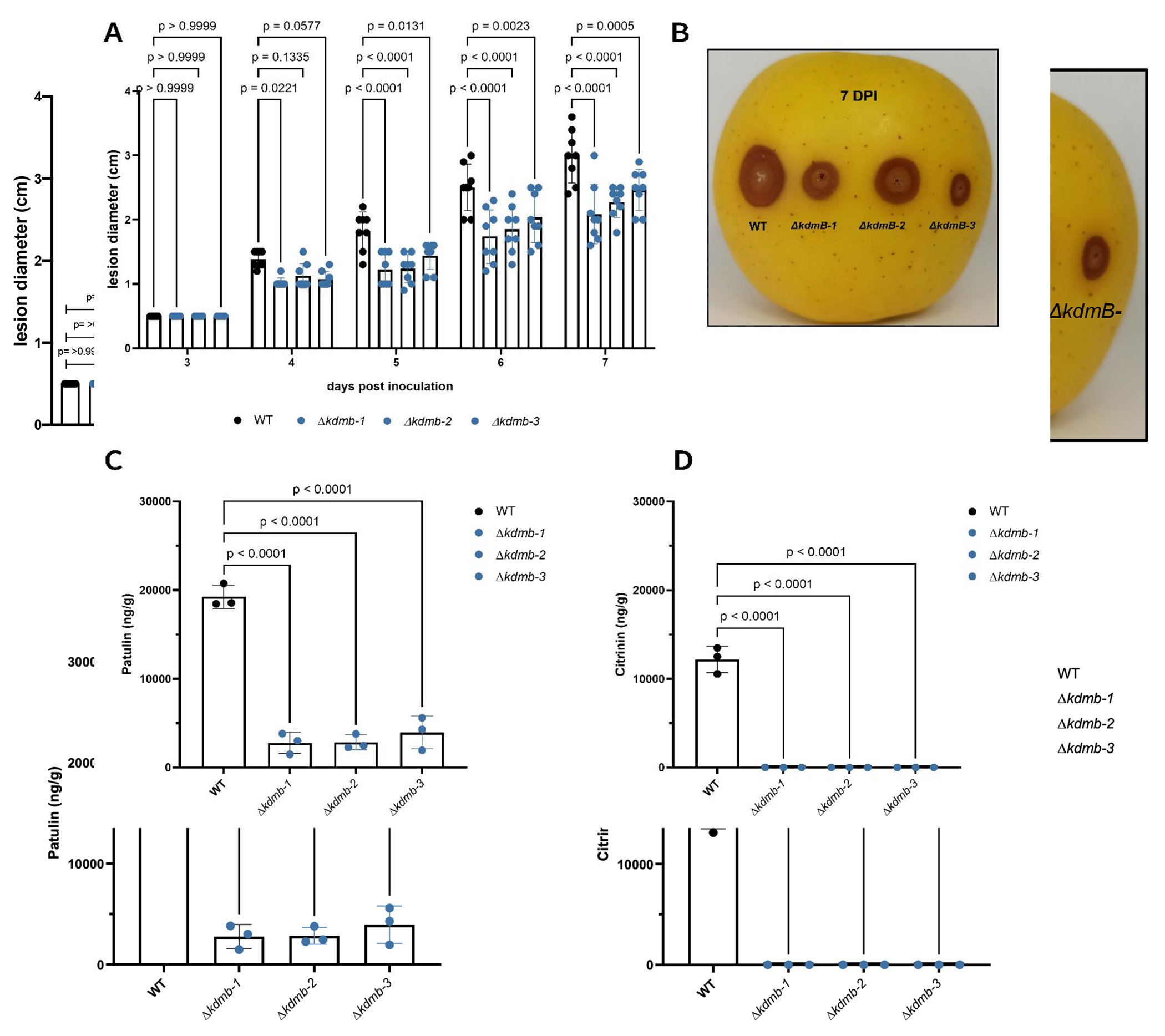
Fig. 3.

Apple lesion development and mycotoxin detection in vivo. (A) Lesion diameters were measured for 7 days upon inoculation with WT or ΔkdmB strains. (B) representative photo of an inoculated apple with the assessed strains. (C) patulin and (D) citrinin were detected from 1g of homogenized infected tissue at 16 dpi. Error bars denote standard deviation of the mean.
