
Fig. 2.
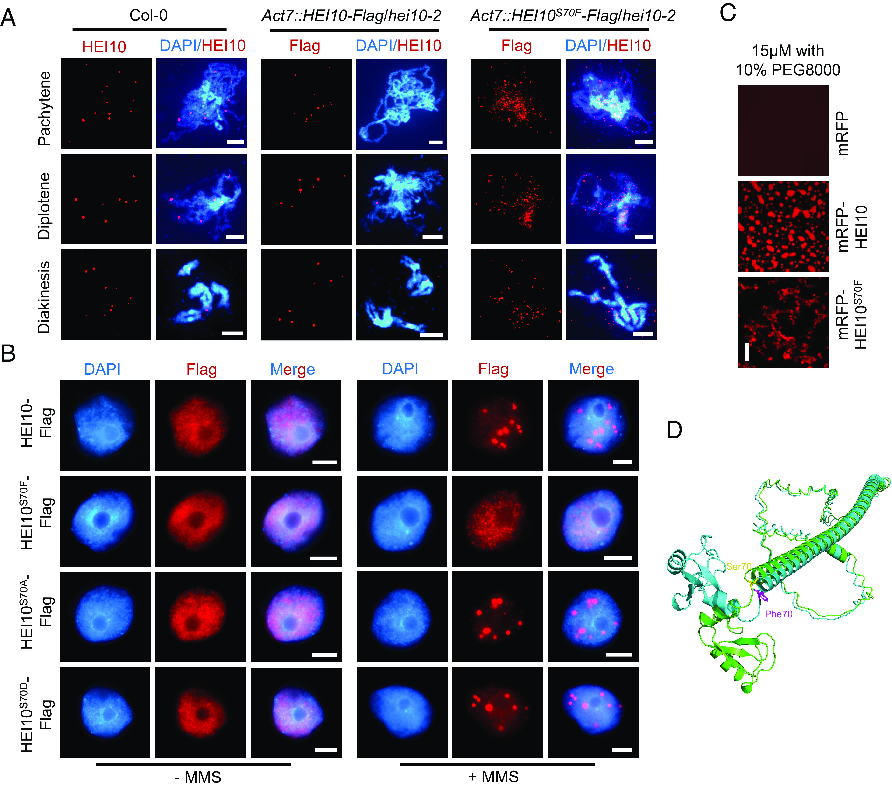
Mutation of S70F affects the phase separation of HEI10. (A) Immunostaining of HEI10 at pachytene, diplotene, and diakinesis in Col-0, HEI10-Flag, and HEI10S70F-Flag transgenic plants. (Scale bar, 5 μm.) (B) Immunostaining of HEI10-Flag and HEI10S70F/A/D-Flag in tobacco nuclei with or without MMS treatment. The leaves are treated with MMS for 1.5 h. HEI10S70A and HEI10S70D simulate the phospho-dead and phospho-mimetic proteins. (Scale bar, 5 μm.) (C) In vitro phase separation assay of mRFP, mRFP-HEI10, and mRFP-HEI10S70F recombinant proteins, with protein concentrations of 15 μM and 10% PEG8000. (Scale bar, 20 μm.) (D) The 3D structures of HEI10 (green) and HEI10S70F (blue), predicted by ESMFold. The Ser70 (yellow) and the Phe70 (purple) residues are shown at the junction between Ring domain and α-helix by stick representation.