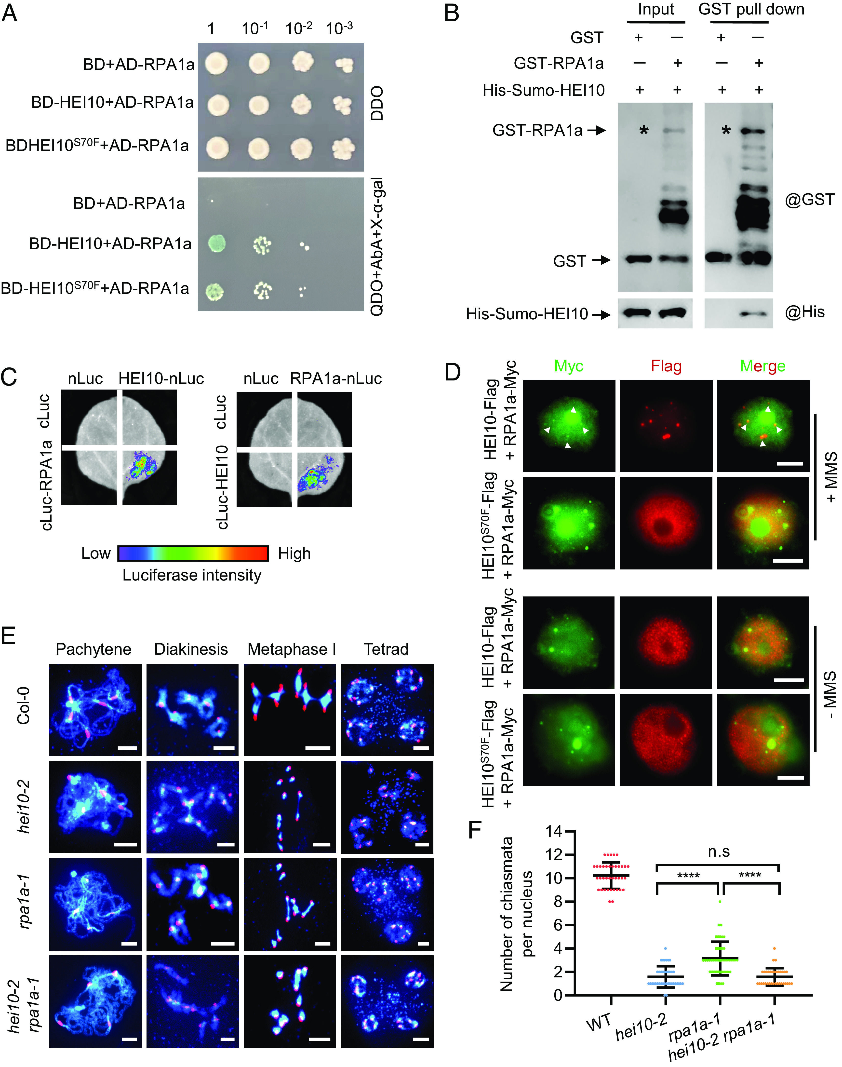
Fig. 3.

HEI10 interacts with RPA1a in vitro and in vivo. (A) Interactions of HEI10 with RPA1a by yeast two-hybrid using gradient dilution. The initial concentration is OD600 ≈ 1, diluted 10-, 100-, and 1,000-fold, spotted on DDO (double synthetic dropout media lacking leucine and tryptophan) and QDO (quadra synthetic dropout media lacking leucine, tryptophan, histidine, and adenine) for 3 d. (B) Validation of interaction between HEI10 and RPA1a using the affinity purification GST pull-down assay. Asterisks indicate the GST-RPA1a band. (C) Split luciferase complementation imaging assay examines the interaction between HEI10 and RPA1a in tobacco cells. cLUC or nLUC are used as controls. (D) Colocalization of HEI10-Flag with RPA1a-Myc in tobacco nuclei. (Scale bar, 5 μm.) White arrows indicate the RPA1a-Myc signals that merged with HEI10-Flag. (E) Meiotic chromosome phenotypes of Col-0, hei10-2, rpa1a-1, and hei10-2 rpa1a-1 using centromere FISH. (Scale bar, 5 μm.) (F) Statistical analysis of the chiasmata at metaphase I in Col-0 and mutants (****P < 0.0001, two-tailed Student's t test).
