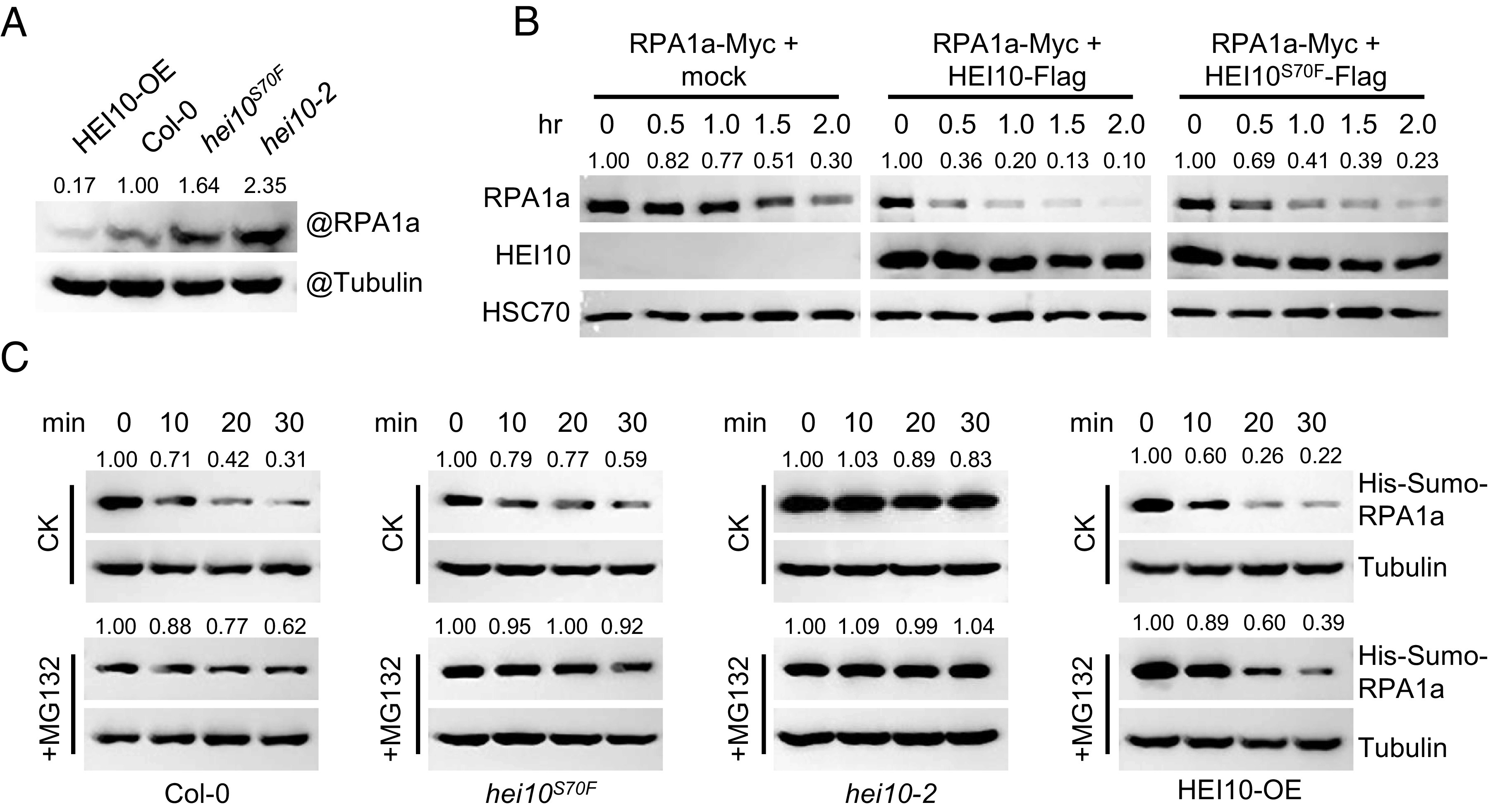
Fig. 5.

HEI10 phase separation affects the degradation of RPA1a. (A) Measurement of endogenous RPA1a protein level in HEI10-OE (Act7::HEI10-Flag/Col-0), Col-0, hei10S70F, and hei10-2 backgrounds with anti-RPA1a antibody. Tubulin is the loading control. (B) Semi-in vivo protein degradation assay, RPA1a proteins extracted from tobacco cells are mixed with HEI10-Flag, HEI10S70F-Flag, and mock control in a volume ratio of 1:2. The mixtures are incubated at room temperature for different time courses. (C) Cell-free degradation assay, recombinant His-Sumo-RPA1a proteins are incubated with equal amount of central inflorescence extractions of HEI10-OE, Col-0, hei10S70F, and hei10-2, and 100 μM cycloheximide (a protein biosynthesis inhibitor) and 10 mM ATP are added into the buffer and the samples are incubated at room temperature. For MG132 treatment, 50 μM MG132 is added. The protein level of His-Sumo-RPA1a is determined with anti-His antibody. Tubulin is the loading control.
