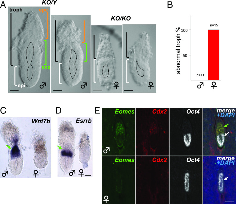
Fig. 1.
Absence of the exe structure in female embryos lacking Rlim. Male/female littermates within subfigures are shown at the same magnification. Sex was determined after image recording. (A) Representative brightfield images of E5.75 embryos. Approximate embryo and lumen domains are indicated. n > 27 each. epc = ectoplacental cone region (brown); exe = extraembryonic ectoderm region (green); troph = trophoblast-derived (black); epi = epiblast (white). (Scale bars: 60 µm.) (B) Quantification of phenotypic appearance. (C and D) Whole embryo in situ hybridization. Lack of exe markers Wnt7b (C) and Esrrb (D) in E5.75 Rlim KO females (n > 10, each). (Scale bars: 60 µm.) (E) IHC of Rlim KO embryonic sections at E5.5 within decidua reveals lack of exe markers Eomes and Cdx2. (Scale bar: 40 µm.) In (C–E), green and white arrows indicate exe and epiblast regions, respectively.

