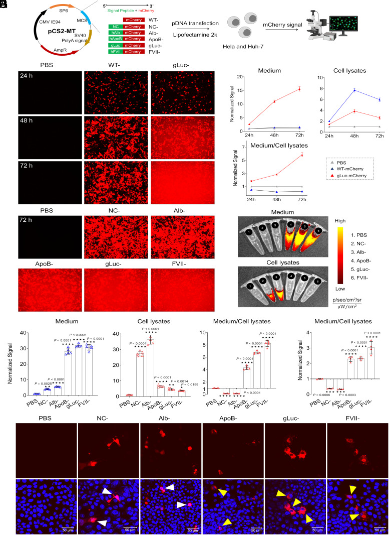
Fig. 1.
Optimal signal peptides (SPs) drive protein secretion effectively following plasmid DNA (pDNA) delivery. (A) Engineering secreted mCherry by including a SP upstream of the coding sequence and screening of optimal SP by pDNA transfection in vitro. (B) Time-dependent mCherry secretion by HeLa cells (exposure time, 1/30 s); mCherry signal was observed clearly in the medium 2 d after treatment with gLuc-mCherry. (C) Quantification of mCherry fluorescence in cell lysates and medium at different time points. (D) SP screening in HeLa cells at 72 h (exposure time, 1/70 s). (E) Cell lysates and medium were imaged and (F) quantified at 72 h. Data is presented as mean ± SEM (n = 5 biologically independent samples). HeLa cells were treated with Lipofectamine 2000 containing pDNA in a 96-well plate (50 ng per well). At noted times points, cells were imaged via confocal microscopy; mCherry signal was quantified by plate reader or captured by IVIS. (G) The functionality of SP in Huh7 cells was confirmed by quantifying mCherry signal in cell lysates and medium at 72 h. Data is presented as mean ± SEM (n = 4 biologically independent samples). (H) A “circle-like” signal (indicated by yellow arrows) was observed in Huh7 cells imaged by confocal microscopy indicative of high efficacy secretion mediated by SP. (Scale bar, 50 µm.) WT, wild type; NC, negative control; Alb, human albumin; ApoB, human apolipoprotein B; gLuc, Gaussia luciferase; FVII, human Factor VII. A two-tailed unpaired t test was used to determine the significance of the comparisons of data indicated in F and G (*P < 0.05; **P < 0.01; ***P < 0.001; and ****P < 0.0001).

