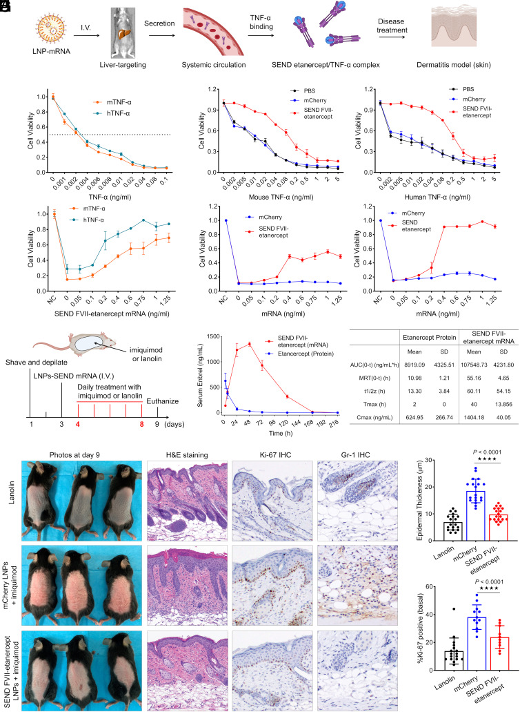
Fig. 4.
SEND FVII-etanercept mRNA LNPs prevent TNF-α induced cell death and achieved a therapeutic benefit in an imiquimod-induced psoriasis model in vivo. (A) Scheme of etanercept (Enbrel) protein production, TNF-α binding, and disease treatment on dermatitis model. (B) Dose-dependent cytotoxicity of human TNF-α (hTNF-α) and mouse TNF-α (mTNF-α) in L929 cells. Cells were incubated 24 h with actinomycin at 1 μg/mL and various concentrations of TNF-α before determining cytotoxicity. (C) L929 cells pretreated by FVII-etanercept mRNA LNPs were resistant to both mouse and human TNF-α. Cells were primed with 80 ng FVII-etanercept mRNA LNPs treatment per well and, 48 h later, were challenged with TNF-α ranging from 0 to 5 ng/mL and a fixed concentration of actinomycin at 1 μg/mL. Twenty-four hours after incubation, cell viability was assessed. (D) Dose-dependent rescue of cell viability following pretreatment by LNPs. Cells were pretreated by FVII-etanercept mRNA LNPs with mRNA doses of 0 to 1.25 ng/mL, after 48 h, cells were challenged by TNF-α with dose of 0.1 ng/mL and fixed actinomycin dose of 1 μg/mL. Twenty-four hours postchallenge, cell viability was measured. (E) Dose-dependent rescue of cell viability after treatment with medium from FVII-etanercept mRNA LNP-treated cells. L929 cells were treated with FVII-etanercept mRNA LNPs with mRNA doses of 0 to 1.25 ng/mL. After 2 d, medium from these cells containing secreted etanercept was collected and transferred into fresh L929 cells, and the cells were challenged by mouse or human TNF-α at a dose of 0.1 ng/mL and actinomycin at 1 μg/mL. Twenty-four hours after media transfer and challenge, cell viability was evaluated. Data are presented as mean ± SEM (n = 5 biologically independent samples) (F) Mice were shaved and depilated on day 1 and subsequently injected with LNPs on day 3. On days 4 to 8, mice were treated with either lanolin or imiquimod to induce dermatitis, and mice were euthanized on day 9. (G) Serum concentration curves for etanercept protein and mRNA following a single I.V. injection at a dose of 0.5 mg/kg native protein or mRNA-LNP formulation. Pharmacokinetic comparison between etanercept protein and mRNA after single dosing. Serum was collected at different time points, and etanercept was detected by ELISA. (H) Images of dorsal skin of lanolin-treated mice (control) and imiquimod-treated mice, which were injected with either mCherry mRNA or FVII-etanercept mRNA formulations. Images of H&E-stained sections, Ki-67 staining, and Gr-1 staining of the dorsal skin of lanolin control and imiquimod-treated mice. (I) Measurement of epidermal thickness in three groups and the percentage of Ki-67+ cells in epidermal basal cells (per 50 cells) quantified from (H). (****P <0.0001).

