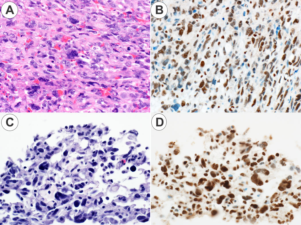
Figure 4. ALT positive angiosarcoma.

Panel 4A. The angiosarcoma case 1 shows a solid, epithelioid growth pattern (original magnification 20X). Panel 4B. The angiosarcoma case 1 is ATRX positive (original magnification 20X). Panel 4C. The angiosarcoma case 2 shows a vasoformative growth pattern (original magnification 30X). Panel 4DB. The angiosarcoma case 2 is ATRX positive (original magnification 30X).
