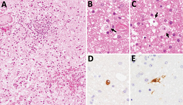
Figure 1.

Brain biopsy from patient (patient 2) with Macacine alphaherpesvirus 1 (herpes B virus) infection, Japan, 2019. A–C) Inflammatory cell infiltration and hemorrhage observed around blood vessels in the cerebellar white matter. Arrows indicate nuclear inclusion bodies (B, C). Hematoxylin and eosin stain. D, E) Immunohistochemical analysis using B virus gB mouse monoclonal (clone 19B6) (D) and an B virus rabbit polyclonal (E) antibodies as the primary antibodies. Original magnification × 200 for all images.
