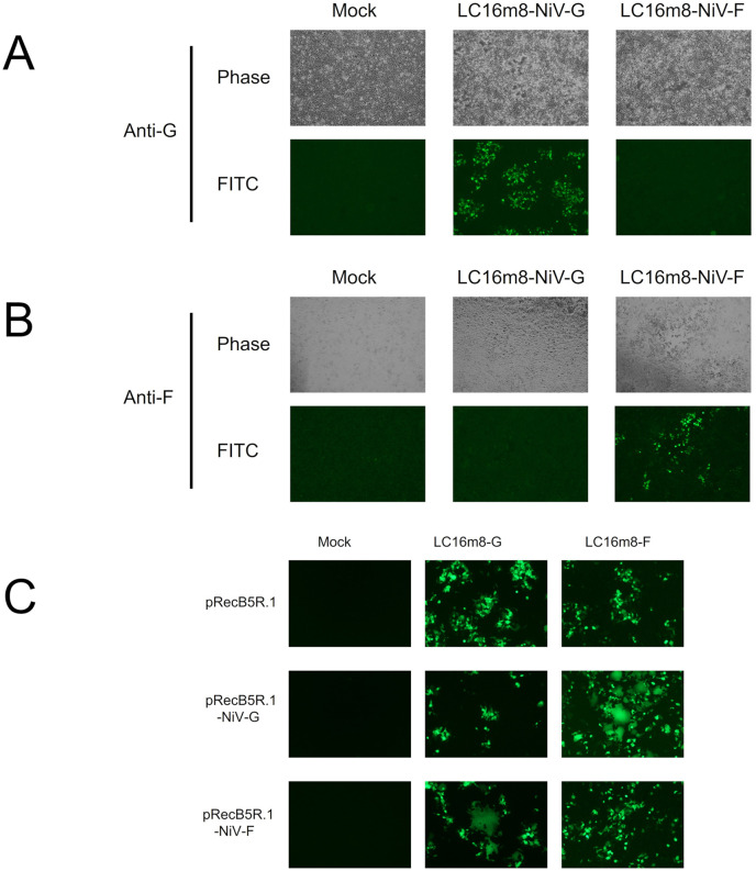
Fig 2. Expression and function of the envelope glycoproteins of NiV.
(A and B) RK-13 cells were infected with LC16m8-G or LC16m8-F at an MOI of 0.01. At 40 hpi, the cells were fixed and stained with a polyclonal antibody against NiV G protein (A) or F protein (B), followed by incubation with Alexa Fluor 488-conjugated anti-rabbit secondary antibody. The stained cells were observed under a phase-contrast or fluorescence microscope. (C) RK-13 cells were transfected with an expression plasmid driven by the vaccinia early and late promoter (pRecB5R.1-NiV-G, pRecB5R.1-NiV-F, or pRecB5R.1). At 24 h posttransfection, the transfectant cells were infected with LC16m8-G or LC16m8-F at an MOI of 0.01. At 24 hpi, the formation of syncytia was observed under a fluorescence microscope.

