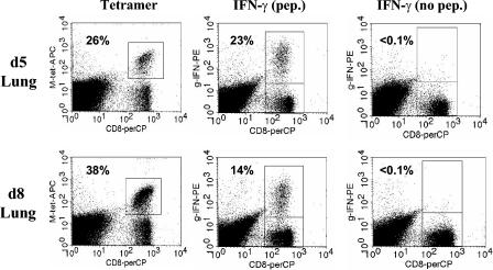
FIG. 7.
Secondary effector cells in the lung become increasingly nonfunctional over time following infection with SV5. BALB/c mice were infected i.n. with 106 PFU of WT rSV5. After d40 postinfection, mice were challenged by i.n. administration of 107 PFU of WT rSV5. Lung cells were either stained with M285-293/Ld tetramer or stimulated with 1 μM M285-293 peptide to assess IFN-γ production as described for Fig. 1. As a negative control, cells were stimulated in the absence of M285-293 peptide. The numbers represent the percentages of CD8+ T cells that were either tetramer positive or IFN-γ+.

