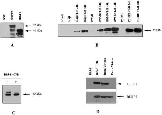
FIG. 2.
(A) Western immunoblot showing the specificity of MAb C1. Samples representing His-BFLF2 and GST-BFLF2 fusion proteins together with GST were run, and the immunoblot was probed with the anti-BFLF2 MAb C1. (B) BFLF2 protein expression in different cell lines, either uninduced or following activation with TPA and sodium butyrate (T/B). (C) Phosphorylation of BFLF2 in induced B95-8 cells. Twenty micrograms of cell lysate was treated with λ-phosphatase (+) or left untreated (−) and analyzed by immunoblotting. (D) BFLF2 is present in intracellular virions. Equal amounts of intracellular and extracellular virion lysates were run together with uninduced and induced B95-8 (T/B) cell extracts. The immunoblot probed with the anti-BFLF2 MAb C1 shows positivity only in intracellular virions and in B95-8+T/B cells. As a control for virion preparation, an antibody specific for a viral capsid antigen (BLRF2) was used.

