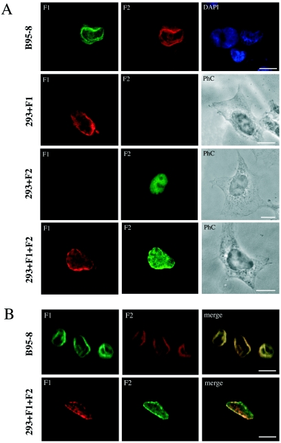
FIG. 6.
Indirect immunofluorescence of B95-8 and 293 cells expressing BFLF2 and BFRF1. (A) Double immunofluorescence of chemically induced B95-8 cells and of 293 cells transfected with CMV-BFRF1 (293+ F1) or CMV-BFLF2 (293+F2) or cotransfected with CMV-BFRF1 and CMV-BFLF2 (293+F1+F2). Bars, 10 μm. PhC, phase contrast. (B) Confocal immunofluorescence of induced B95-8 cells and cotransfected 293 cells, as described for panel A. Yellow staining in the merged images shows colocalization of the two signals. Bars, 10 μm.

