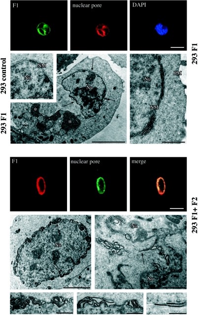
FIG. 8.
Immunofluorescence and electron microscopic analyses of the nuclear membrane structure in transfected 293 cells. Upper panels: CMV-BFRF1-transfected cells (293 F1). Lower panels: CMV-BFRF1- and CMV-BFLF2-cotransfected cells (293 F1+F2). Double-immunofluorescence staining with anti-nuclear pore complex and anti-BFRF1 antibodies reveals an irregular concentration of the two signals in both transfected and cotransfected cells (bar, 10 μm). Electron microscopic analysis of 293 F1 cells reveals focal multilayering of the nuclear membrane (NM) (arrows in the left panel and detail in the right panel), which is not observed in untransfected cells (293 control). Asterisks indicate cytoplasmic membrane structures possibly corresponding to aggregates of endoplasmic reticulum cisternae (asterisks in the left panel). Nu, nucleus; PM, plasma membrane. (Bars, in sequential order: 1, 5, and 1 μm.) Parallel electron microscopic analysis of 293 F1+F2 cells shows a highly irregular structure of the nuclear membrane, with more pronounced multilayering (enlargements of the areas indicated by arrows are shown in the lower panels). (Bars, in sequential order: 5, 2, 1, 1, and 0.5 μm.)

