FIG. 7.
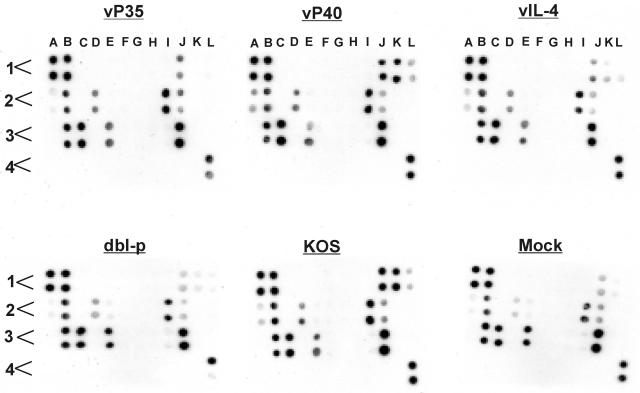
Detection of cytokines and chemokines secreted by lymphocytes of mice immunized with recombinant viruses. Spleens from immunized mice were harvested 3 weeks after the third immunization. Single cell suspensions of T cells were prepared and subjected to in vitro stimulation for 72 h with 5 PFU of UV-inactivated McKrae/cell as described in Materials and Methods. The presence of cytokines and chemokines in the supernatant was determined by an antibody microarray. Each cytokine or chemokine is presented in duplicate, and each panel represents the pattern of cytokine and chemokine expression for vP35, vP40, vIL-4, dbl-p, KOS, or mock-immunized mouse. The order of cytokines and chemokines on each template are as follows: A1 (positive control), A2 (IL-5), A3 (MCP-5, monocyte chemotactic protein 5), A4 (blank), B1 (positive control), B2 (IL-6), B3 (MIP-1α), B4 (blank), C1 (negative control), C2 (IL-9), C3 (MIP-2), C4 (blank), D1 (negative control), D2 (IL-10), D3 (MIP-3β), D4 (blank), E1 (6Ckine), E2 (IL-12p40), E3 (RANTES), E4 (blank), F1 (cutaneous T-cell-attracting chemokine or CCL27), F2 (IL-12p70), F3 (stem cell factor), F4 (blank), G1 (eotaxin), G2 (IL-13), G3 (soluble TNF receptor type I), G4 (blank), H1 (granulocyte-colony stimulating factor), H2 (IL-17), H3 (thymus and activation regulated chemokine), H4 (blank), I1 (granulocyte-macrophage colony-stimulating factor), I2 (IFN-γ), I3 (tissue inhibitor of metalloproteinase 1), I4 (blank), J1 (IL-2), J2 (KC), J3 (TNF-α), J4 (blank), K1 (IL-3), K2 (leptin), K3 (thrombopoietin), K4 (blank), L1 (IL-4), L2 (monocyte chemoattractant protein 1), L3 (vascular endothelial growth factor), and L4 (positive control). The results are repeated twice.

