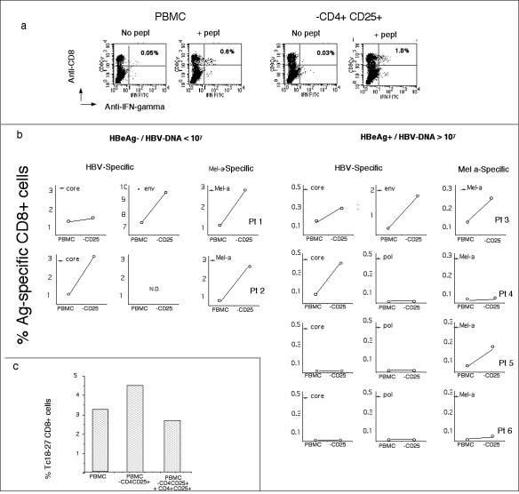
FIG. 3.
Depletion of CD4+ CD25+ cells enhances the expansion of CD8+ T cells. All experiments were performed on short-term lines derived from PBMC or CD4+ CD25+-depleted PBMC stimulated with HBV and Melan-A peptides. The frequency of peptide-specific IFN-γ-producing CD8+ cells was tested with ICS after 8 days of in vitro expansion. (a) Dot plots show the frequency of env183-191-specific CD8 cells obtained in the different expansion conditions (PBMC or CD4+ CD25+-depleted PBMC). IFN-γ-producing CD8+ cells specific for env183-191 were quantified with ICS by stimulating cells for 6 h with env183-191 peptide (+ pept). Negative controls are cells not stimulated with peptide (No pept). (b) Frequencies of IFN-γ-producing CD8+ T cells specific for HBV or Melan-A peptides and obtained in the indicated patients. Note that the scale on the y axis differs in the panels. (c) CD4+ CD25+ cells regulate CD8 expansion. Purified CD4+ CD25+ cells were added back into PBMC of patient 1 depleted of CD25+ CD4+ cells (PBMC/CD4+ CD25+ ratio = 20/1). Cells were stimulated with peptide core18-27. The frequency of core18-27 CD8 was analyzed after 10 days of in vitro expansion. Bars indicate the frequency of expanded core18-27 CD8+ cells out of total CD8+ cells calculated by staining cells with Tc18-27 tetramers and anti-CD8. The experiment was repeated twice in patient 1 with similar results.

