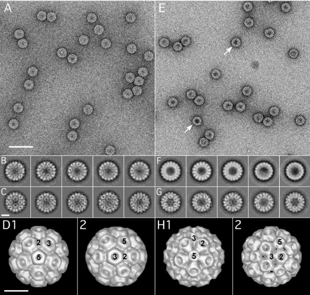
FIG. 2.
Electron microscopy and image analysis of WT CCMV and C3/Δ919 virions. Electron micrographic images of purified virions of WT CCMV (A to D) and Δ919 (E to F). Images shown in panels A and E represent typical regions of electron micrographs of WT and Δ919 mutant virus particles, respectively, negatively stained with 1% uranyl acetate. Images shown in panels B and F represent class sums after the alignment by classification step. Uniform size was observed for the WT (B), whereas for the Δ919 mutant, two particle sizes were observed In panel F, particles shown in the first three images from the left are WT size, while particles shown in the remaining two images are approximately 1.5 nm smaller in diameter than those of the WT. Images C and G show averages after the final MRA for WT and the larger of the two Δ919 populations, respectively. Images D and H represent the final three-dimensional reconstructions for the WT and the larger Δ919 particles, respectively. The positions of the five-, three-, and twofold symmetry axes are indicated. Scale bars are 60 nm (A and E) and 10 nm (B through H). The resolution of the final models was estimated to 25 Å by the Fourier shell criterion with a cutoff of 0.5 for both WT and mutant virions.

