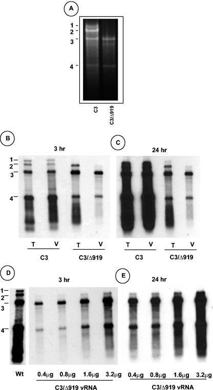
FIG. 3.
Analysis of virion RNA content of C3/Δ919 virions. (A) Native agarose gel analysis of viral RNA. RNA isolated from purified virion preparations of WT C3 or C3/Δ919 was subjected to electrophoresis in 1% agarose and stained with ethidium bromide. (B) Northern blot analysis of total nucleic acid (T) and virion RNA (V) preparations recovered from symptomatic cowpea leaves inoculated with WT C1 and C2 and either C3 or C3/Δ919. Approximately 5 μg of total nucleic acid and 200 ng of virion RNA were denatured with formamide-formaldehyde and subjected to 1.5% agarose gel electrophoresis prior to vacuum blotting to a nylon membrane. The blot was hybridized with a 32P-labeled riboprobe complementary to the commonly shared 3′ noncoding region of CCMV RNAs. The autoradiograph shown in panel C represents a longer exposure image of panel B. (D) Concentrations of C3/Δ919 virion RNA ranging between 0.4 to 3.2 μg per ml were subjected to Northern hybridization as described above. The autoradiograph shown in panel E represents a longer exposure image of panel D. The positions of four CCMV RNAs are shown to the left.

