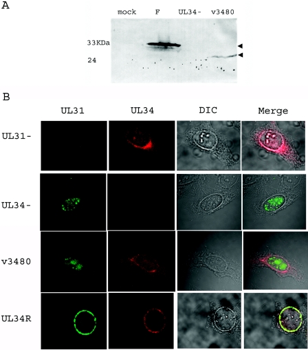
FIG. 5.
(A) Scanned digital image of immunoblot of cell lysates probed with an antibody to pUL34. Hep2 cells were either mock infected or infected with wild-type HSV-1(F), the UL34 deletion virus, or the mutant virus v3480. The cells were lysed, and associated proteins were denatured, electrophoretically separated, transferred to nitrocellulose, and probed with a pUL34-specific antibody. (B) Confocal images of the localization of pUL31 and pUL34 in infected cells. Hep2 cells were infected with the UL31 deletion virus (UL31−), the UL34 deletion virus (UL34−), the mutant virus v3480, or the UL34 repair virus (UL34R) for 16 h and then fixed with ice-cold methanol at −20°C. The cells were then stained with pUL31- and pUL34-specific rabbit and pUL34-specific chicken antibodies, and bound antibodies were detected with a FITC-conjugated donkey anti-rabbit antibody and a Texas Red-conjugated donkey anti-chicken antibody. Differential interference contrast (DIC) images were collected separately. The separately collected channels of fluorescent and DIC images were coprojected in the rightmost column. The intensity of the red and green channels was increased in the merged image to allow comparison with light-refracting features.

