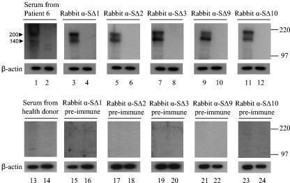
FIG. 2.
Western blot analysis for detection of the S protein. Cos7 cells were transfected with plasmid pKT-S (lanes 1, 3, 5, 7, 9, 11, 13, 15, 17, 19, 21, and 23) or with a plasmid without an insert as a negative control (lanes 2, 4, 6, 8, 10, 12, 14, 16, 18, 20, 22, and 24). Cell lysates were separated in 10% PAGE gel. Western blotting was performed with the antisera, preimmune sera, and control serum indicated on top of each gel. Arrowheads (left), molecular masses (in kilodaltons) of specific S proteins. Membranes were reprobed with mouse anti-β-actin as the loading control. High-range Rainbow molecular weight markers (right; Amersham) were used. α, anti.

