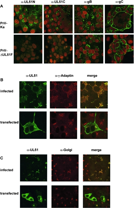
FIG. 2.
Subcellular localization of PrV UL51. (A) RK13 cells were infected with PrV-Ka or PrV-ΔUL51F, fixed 16 h p.i., and incubated with UL51-specific antisera (α-UL51N, α-UL51C) or monoclonal antibodies against gB (A20-c26) or gC (B16-c8). Fluorescence of Alexa 488-conjugated anti-rabbit or anti-mouse antibodies (green) and of propidium iodide-stained chromatin (red) was analyzed in a confocal laser scan microscope. (B and C) RK13 cells were infected with PrV-Ka or transfected with plasmid pcDNA-UL51 and then incubated with the anti-UL51 serum (green fluorescence) and a monoclonal antibody against γ-Adaptin (panel B, red fluorescence) or an unspecified Golgi marker (panel C, red fluorescence) (16). Columns labeled “merge” depict the merger of the green and red fluorescence.

