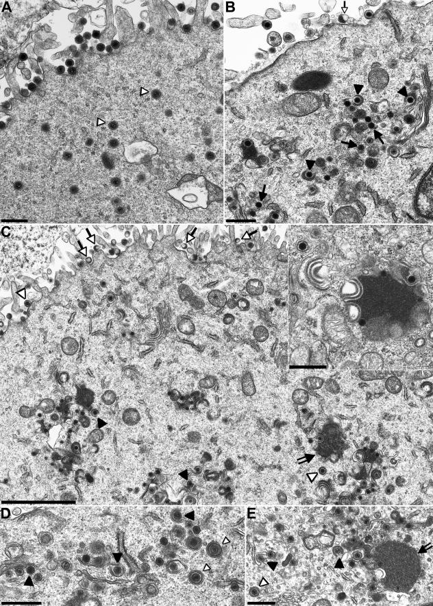
FIG. 6.
Transmission electron microscopy of infected cells. RK13 (A through C), RK13-UL11 (D), or RK13-UL51 (E) cells were infected with PrV-Ka (A), PrV-ΔUL51F (B), or PrV-ΔUL51F/11G (C through E) at an MOI of 1 and fixed 14 h p.i. Arrows point to intracytoplasmic nucleocapsids, closed triangles point to nucleocapsids undergoing secondary envelopment, and open triangles point to enveloped virions. Open arrows indicate L-particles. In PrV-ΔUL51F/11G-infected RK13 or RK13-UL51 cells, intracytoplasmic nucleocapsids were sometimes observed in close contact with tegument and distorted intracytoplasmic membranes (panels C and E, double-shafted arrows; also panel C, inset) as previously described for a PrV UL11 deletion mutant (23, 24), whereas infected RK13-UL11 cells exhibited an increased number of nucleocapsids in the process of secondary envelopment as observed in RK13 cells infected by PrV-ΔUL51F. Bars, 500 nm (A, B, C inset, D, and E) and 2 μm (C).

