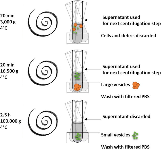
Fig. 1.
Schematic figure of isolation of large and small EVs. EV isolates were prepared from 8 mL serum-free medium after a 24-h incubation on each cell line in a T75 flaks with full confluency. Medium was collected and centrifuged at 3000 g for 20 min at 4 °C, to ensure the removal of aggregates and apoptotic bodies. The supernatants containing the EVs were collected to a clean tube and centrifuged at 16,500 g to pellet large EVs. The supernatant was collected to clean tubes and ultracentrifuged at 100,000 g for 2.5 h at 4 °C to pellet small EVs. Both large- and small EV pellets were washed in 0.2 micron filtered PBS and recentrifuged as described earlier. The EV-enriched pellets were resuspended in 100 µL filtered PBS and used as a fresh sample or stored at -80 °C for further analysis

