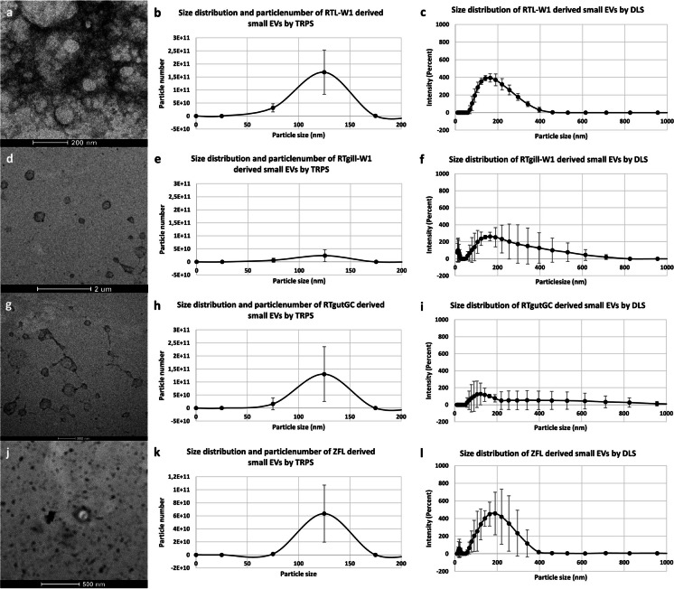
Fig. 3.
Representative images of the size distribution and morphology of small extracellular vesicles isolated from piscine cell lines. TEM images of the RTL-W1 (a); RTgill-W1 (d); RTGC (g) and ZFL (j) cell line derived small EVs. Size distribution and particle number of RTL-W1 (b); RTgill-W1 (e), RTgutGC (h) and ZFL (k) cell line derived small EVs measured by TRPS. Size distribution of RTL-W1 (c); RTgill-W1 (f); RTgutGC (i) and ZFL (l) cell line derived small EVs measured by DLS

