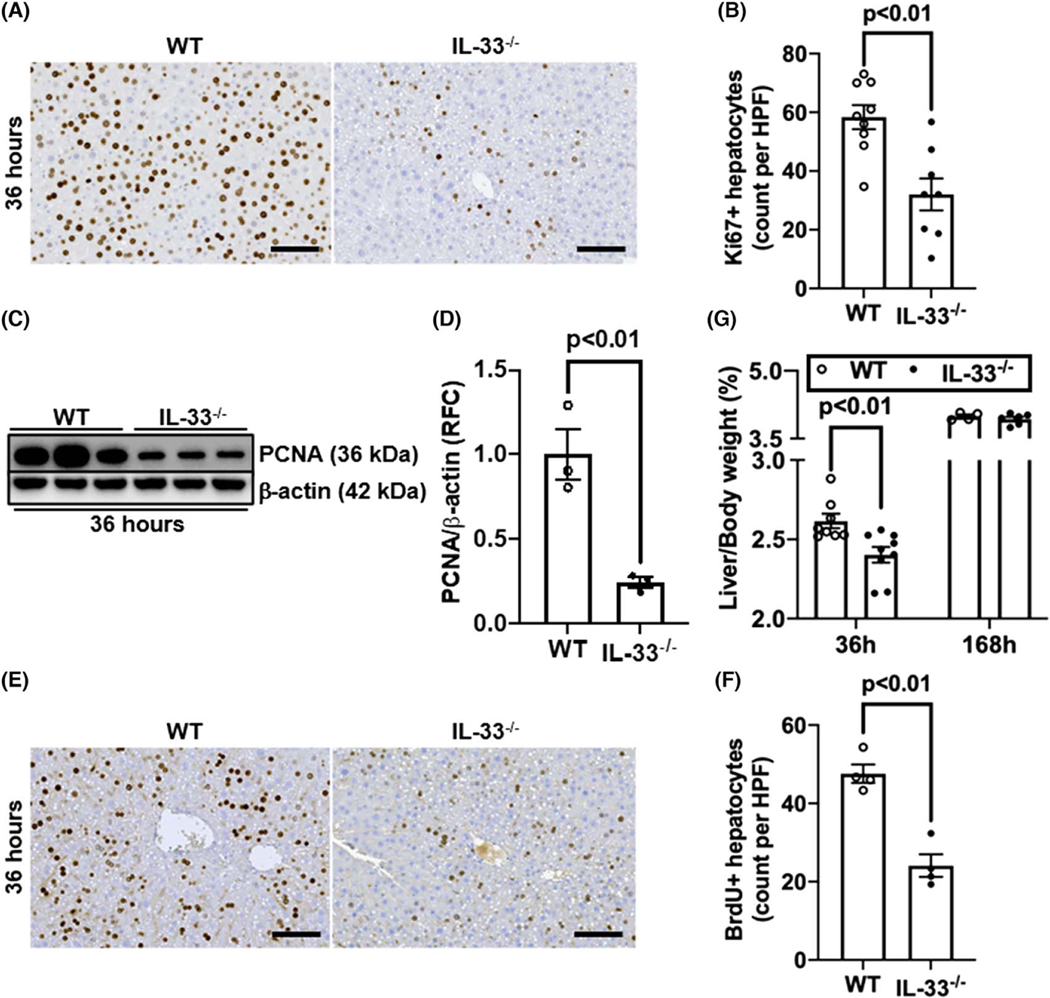
FIGURE 1.
IL-33 deletion delays liver regeneration after partial hepatectomy (PHx). Wild-type (WT) and IL-33−/− mice were subjected to PHx. (A,B) Ki67 immunostaining was performed in liver sections collected at 36 h after PHx (scale bar: 100 μm). Ki67-positive hepatocytes were quantified (n = 8-9/group). (C,D) Proliferating cell nuclear antigen (PCNA) levels in liver tissues at 36 h after PHx were measured by immuno-blotting and the ratios of PCNA/β-actin were quantified (n = 3/group). (E,F) Bromodeoxyurdine (BrdU) incorporation was performed in liver sections at 36 h after PHx (scale bar: 100 μm). BrdU positive hepatocytes were quantified (n = 4/group). (G) The ratios of liver/body weight were analyzed at 36 h and 168 h after PHx (n = 4–9/group). Two-tailed unpaired Student’s t test was performed in (B), (D), and (F). Two-way ANOVA was performed in (G). HPF, high-power field; RFC, relative fold change

