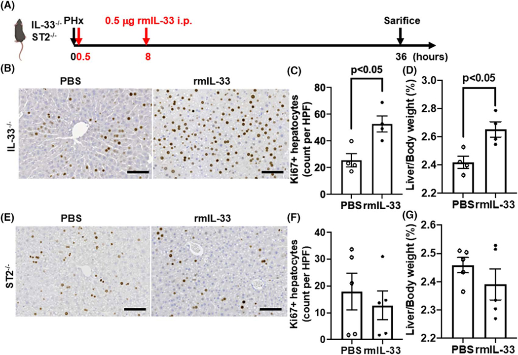
FIGURE 3.
Exogenous IL-33 promotes liver regeneration in IL-33−/− mice after PHx. (A) Strategy of exogenous IL-33 treatments in IL-33−/− and ST2−/− mice after PHx. (B,C) Ki67 immunostaining was performed in liver sections of IL-33−/− mice with PBS or recombinant murine IL-33 (rmIL-33) treatment at 36 h after PHx (scale bar: 100 μm). Ki67-positive hepatocytes were quantified (n = 4/group). (D) The ratios of liver/body weight were analyzed at 36 h after PHx (n = 4/group). (E,F) Ki67 immunostaining was performed in liver sections of ST2−/− mice with PBS or rmIL-33 treatment at 36 h after PHx (scale bar: 100 μm). Ki67-positive hepatocytes were quantified (n = 5/group). (G) The ratios of liver/body weight were analyzed at 36 h after PHx (n = 5/group). Two-tailed unpaired Student’s t test was performed in (C), (D), (F), and (G)

