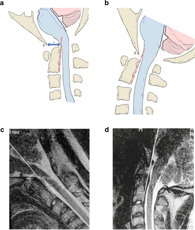
Fig. 2.
a Pathological translation of the basion with respect to the odontoid. Normally, between flexion and extension, the basion (b) pivots over the odontoid with < 2 mm of translation. In Fig. 2a, the cervical spine is in flexion, and the basion has translated anteriorly causing a bend in the brainstem. Note the BAI (*)—the interval measured from the basion to the posterior axial line (dashed line) is greater than the width of the spinal cord. b The cervical spine in extension shows straightening of the brainstem and a upper spinal cord and a shortened BAI. The change in BAI represents a pathological translation of the basion with respect to the spine. c MRI, upright, (dynamic), mid-sagittal, flexion view, T2 weighted (0.6 Tesla, Fonar Corp). The basion axis interval is 12 mm. d MRI, upright, (dynamic), mid-sagittal, extension view, T2 weighted (0.6 Tesla, Fonar Corp). The basion axis interval is 5 mm. Therefore, the BAI in flexion (12 mm) minus the BAI in extension (5 mm) represents a pathological translation of 7 mm

