Figure 1.
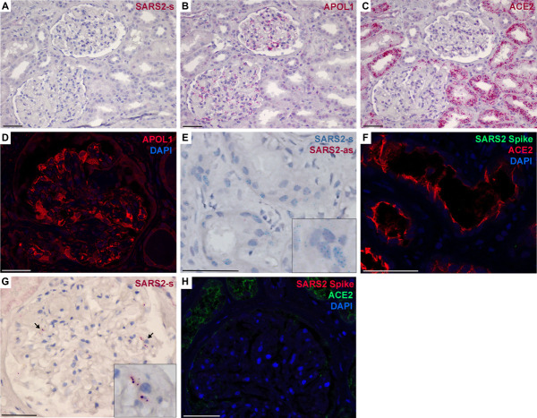
SARS-CoV-2 was rarely detected in human kidney tissue. Representative images of kidney biopsy and autopsy tissue for ACE2, APOL1, and SARS2 sense (s) and antisense (as) RNA by in situ hybridization and ACE2, APOL1 and the viral capsid spike protein by immunofluorescence. (A–C) Serial sections of a kidney biopsy that was negative for SARS2 RNA (A), whereas RNA for APOL1 (B) was evident in podocytes, immune cells, and glomerular and peritubular endothelia, and RNA for ACE2 (C) was abundant in proximal tubules. (D) The detected APOL1 RNA in glomeruli also reflected expression of APOL1 protein by immunofluorescence. (E and F) A single kidney biopsy (one of 26) had a region of SARS2-s RNA positivity in tubules that could not be ruled out as artifact; however, this region was negative for SARS2-as RNA and the viral spike protein (F), indicating a lack of viral replication or intact viral particles. (F) Region of positivity shown in (E) was negative for SARS2 spike protein, although ACE2 was abundant. (G) A single kidney autopsy sample (one of 30) had rare evidence of SARS2-s positivity in glomeruli (arrows, magnification in inset). (H) Autopsy specimen shown in (G) was negative for the SARS2 spike protein. Scale bar=50 μm. APOL1, apolipoprotein L1; SARS-CoV-2, severe acute respiratory syndrome coronavirus 2.

