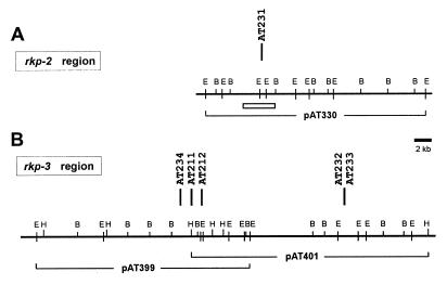
FIG. 2.
Physical-genetic map of the rkp-2 (A) and rkp-3 (B) regions of S. meliloti. Vertical bars represent the Tn5 insertions (with strain numbers) identified in the phage φ16-3-resistant mutants. All mutants were defective in KPS production (see Fig. 1). The physical map was constructed by EcoRI (E), BamHI (B), and HindIII (H) restriction enzymes. Solid lines under the physical map show the genomic DNA insert and the number of the complementing cosmid clones. In panel A, a rectangle indicates the region where the DNA sequence was established. In panel B, mutations Tn5-AT211 and Tn5-AT212 were complemented by both cosmid clones.

