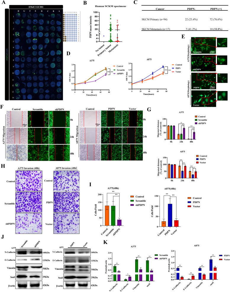
Fig. 1.
PDPN is upregulated in melanoma, and is associated with the proliferation and metastasis of melanoma. A Human melanoma tissue microarrays named HMelC112CD01 consisting of nontumor melanoma sample (n = 1), primary melanoma samples (n = 94), and melanoma distant metastasis samples (n = 17). B Human melanoma samples were subjected to immunofluorescence for PDPN with quantitative analyses. C Statistically PDPN expression with different melanoma samples. D PDPN expression correlated with melanoma cell lines (A375, A875) proliferation as measured by CCK8 assay. E Images demonstrated morphological changes after stable knockdown of PDPN in A375 cells and high expression of PDPN in A875 cells. The power field scale bar, 100 μm. F, G Wound healing assays were performed for the migration capability of A375 cells stably knockdown PDPN or A875 cells with PDPN overexpression, and statistical analysis was performed to determine the migrated distance. The power field scale bar, 100 μm. H, I Transwell analysis was performed to quantify the invasive ability of PDPN knockdown or overexpression cells, and statistical analysis was performed to determine the invasion of cells. The power field scale bar, 100 μm. J, K Western blot analysis was performed to identify the effects of PDPN on the EMT markers E-Cadherin, N-Cadherin, vimentin, and snail. β-actin as an internal control was used. The statistical analysis was performed to quantify the relative protein levels. Data are presented as mean ± SD. *p < 0.05, **p < 0.01, ***p < 0.001 versus control

