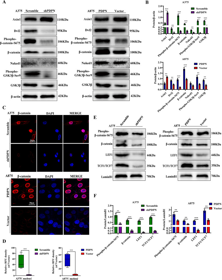
Fig. 2.
Effect of PDPN on the Wnt/β-Catenin signaling pathway in melanoma cells. A Western blot analysis was performed to detect the Wnt/β-catenin signaling-related proteins. β-actin as an internal control was used. B Statistical analysis was performed to quantify the relative protein levels. C Immunofluorescence staining was conducted to quantify PDPN effects on the nuclear β-catenin intensity in melanoma cell lines. The power field scale bar, 20 μm. D Statistical analysis was performed to quantify the immunofluorescence intensity of nuclear β-catenin in A875 cells. E Western blot assay was also performed on nuclear proteins related to the Wnt/β-catenin signaling pathway including β-catenin, phospho-β-catenin, LEF1, and TCF1/TCF7. The LaminB1 was used as an internal control for nuclear proteins. F Statistical analysis was performed to quantify the relative protein levels. Data are presented as mean ± SD. *p < 0.05, **p < 0.01, ***p < 0.001 versus control

