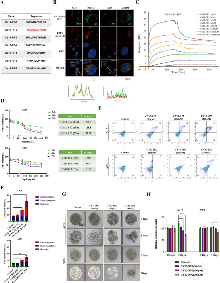
Fig. 3.
Biopanning of PDPN antagonist peptides. A A summary table of peptide sequences selected from the fourth round phage biopanning of PDPN peptides using a phage display library kit. B Immunofluorescence staining was performed to quantify the colocalization of CY12-RP2 and PDPN protein in melanoma cell lines (A375, A875). The power field scale bar, 20 μm. C CY12-RP2 (100, 20, 4.0, 0.8, 0.16, 0.032, 0.0064, 0.00128 μM) dose-dependent binding to PDPN. D Suppression efficiency of melanoma cells growth by CY12-RP2 was measured by CCK8 assay at various concentrations for 24, 48 or 72 h, respectively. E, F The A375 and A875 cells were treated with various concentrations of CY12-RP2 (0, 50, 100, 200 μM) for 48 h and analyzed using the Annexin V/PI staining flow cytometry (E), and statistical analysis was performed to quantify the apoptosis rates (F). G, H The 3D cellular spheres were treated with the set concentrations of CY12-RP2 for 5 days and cell morphology was assessed (G), relative spheroid diameter was measured (H). Data are presented as mean ± SD. *p < 0.05, **p < 0.01, ***p < 0.001 versus control. The power field scale bar, 100 μm

