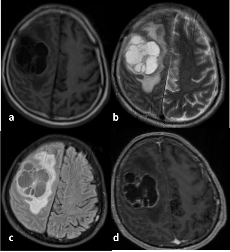
Fig. 6.

a Axial T1-weighted, (b) T2-weighted, (c) FLAIR, and (d) post-contrast administration T1-weighted MR images from a 60-year-old male with a Glioblastoma WHO CNS Grade 4, IDH-wildtype. There is a distinct and relatively thick area of enhancement on the edge of the necrotic area, with a cumulative size of about a third of the total tumor area. The abnormal FLAIR area appears slightly larger than the pathological intensity on the T1-weighted image. This glioma carries the IDH-wildtype marker. The tumor almost has no non-enhancing areas as it primarily consists of necrotic areas with a solid enhancing area at its edge. The longest diameter of the tumor size is relatively not too large, and the tumor's edema area seems limited to one hemisphere. This tumor is also MGMT-unmethylated
