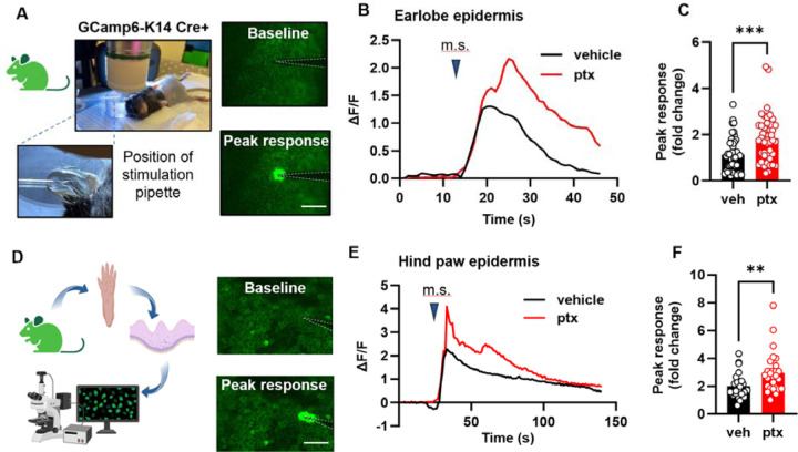
Fig. 2. Paclitaxel treatment sensitizes the mouse epidermis to mechanical stimulation.
(A) Visual representation and example fluorescent images of the in vivo mechanical calcium imaging setup used on the earlobe skin of GCamp6-K14 Cre+ mice. (B) Example traces illustrating calcium responses induced by mechanical stimulation (m.s.) in earlobe skin. (C) Comparative analysis of peak calcium responses to mechanical stimuli of earlobe skin in paclitaxel (ptx) and vehicle-treated mice. Data was sourced from 4 mice per group, with 5–15 recordings per mouse (totaling 45–46 recordings per treatment group). (D) Diagram showcasing the experimental approach for in situ calcium imaging on paw keratinocytes. Hindpaw skin from GCamp6-K14 Cre+ mice underwent dissection, with the epidermis subsequently separated from the dermis for imaging purposes (left). Fluorescence images of the paw epidermis both at baseline and immediately following mechanical stimulation (right). (E) Representative traces displaying calcium responses induced by mechanical stimulation (m.s.) in the hind paw epidermis. (F) Comparative analysis of peak calcium responses to mechanical stimuli of hind paw epidermis of paclitaxel (ptx) and vehicle-treated mice (right). Data comprises recordings from 5 mice per group, with 5 recordings taken per mouse (yielding 25 recordings for each treatment group). Scales are 50 μm. Statistical analysis was performed using Student’s T test; **P < 0.01, and ***P < 0.001. Error bars represent SE. Schematic created with BioRender.com.

