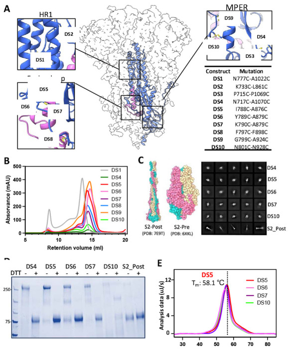
Figure 1. Characterization of covalently-linked SARS-CoV-2 S2 variants.

(A) Side view of the trimeric SARS-CoV-2 spike (silhouette) with a schematic representation of a monomer of S2 (PDB:6XKL) in ribbon diagram. The fusion peptide (FP) is colored in magenta. Each inset corresponds to the location of the computationally beneficial introduction of cysteines to form disulfide bonds. The position of each substitution is shown in the table. (B) Size-exclusion chromatography (SEC) traces comparing S2 purity post-purification. (C) Atomic diagram of the post- and pre-fusion conformations with each monomer colored differently. Representative negative-stain electron 2D classes of the S2-prefusion candidates DS4, DS5, DS6, DS7, and DS10, showing homogenous well-formed trimer populations, and S2 post-fusion construct from wild-type S2. (D) Non-reducing/Reducing SDS-PAGE of constructs shown in (C). (E) Differential Scanning Calorimetry (DSC) analysis of spike thermostability.
