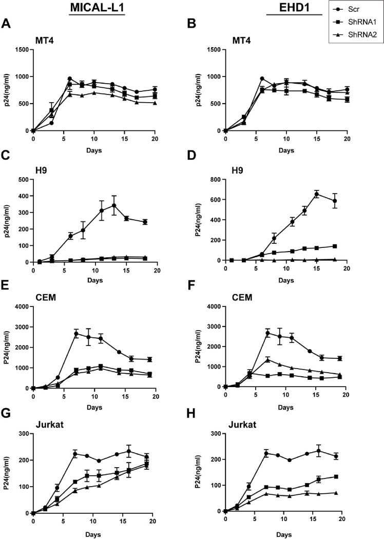
Figure 6. Depletion of MICAL-L1 or EHD1 results in replication defects in a cell type-specific manner.
Cells were infected with NL4–3 and maintained for 3 weeks with intermittent sampling to assess virus release using p24 antigen assay. Growth curves following depletion of MICAL-L1 (left) or EHD1 (right) in permissive MT-4 cells (A,B), nonpermissive H9 cells (C,D), nonpermissive CEM cells (E,F) and nonpermissive Jurkat cells (G,H).

