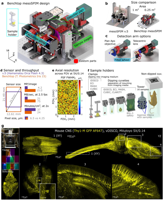
Fig 1. The Benchtop mesoSPIM design and an application example.
a, CAD model of the microscope with main modules labelled: excitation arms, detection path, sample stages, and sample holder. Modified or custom-made parts are red. b, Size comparison between mesoSPIM v.5 and Benchtop systems. c, Detection arm can be equipped with a plan apochromatic objective (2x-20x magnification, tube lens not shown) or a telecentric lens (0.9x-2x) depending on the application, with a corresponding filter wheel and set of filters. The detection arm is mounted on a focusing stage. d, Comparison of sensor size, pixel size, pixel count per image, and imaging throughput between v.5 (Hamamatsu Orca Flash4.3 camera) and Benchtop (Teledyne Photometrics Iris 15). e, The Benchtop mesoSPIM axial resolution in ASLM mode across the field of view, at magnification 5x and NA 0.14. The full width at half-maximum of point-spread function along the z-axis (FWHMZ) is color-coded from 0 to 5 μm. The resolution was measured with 0.2 μm fluorescent beads embedded in agarose immersed high-index medium (RI=1.52). f, CAD models of custom 3D printed sample holders that accommodate samples from 3 mm to 75 mm. g, Example of a multi-tile imaging of a mouse CNS (Thy1-GFP line M, Atto647N, cleared with vDISCO) at 5x magnification. The workflow consists of sample mounting, tiled acquisition, and stitching. This cm-scale sample was imaged at about 3 μm resolution that shows axonal and dendritic arborizations of long-projecting neurons: motor axons in the spinal cord (inset 1), Purkinje cells (inset 2), pyramidal cells in cortex and hippocampus (inset 3), pyramidal cells in the prefrontal cortex (inset 4). Maximum intensity projections (MIPs) over multiple planes spanning 500 μm (insets 1 and 2), 1000 μm (insets 3 and 4), or the entire volume (YZ projection of the brain) are shown. Gamma correction (1.5 in Imaris) was applied.

