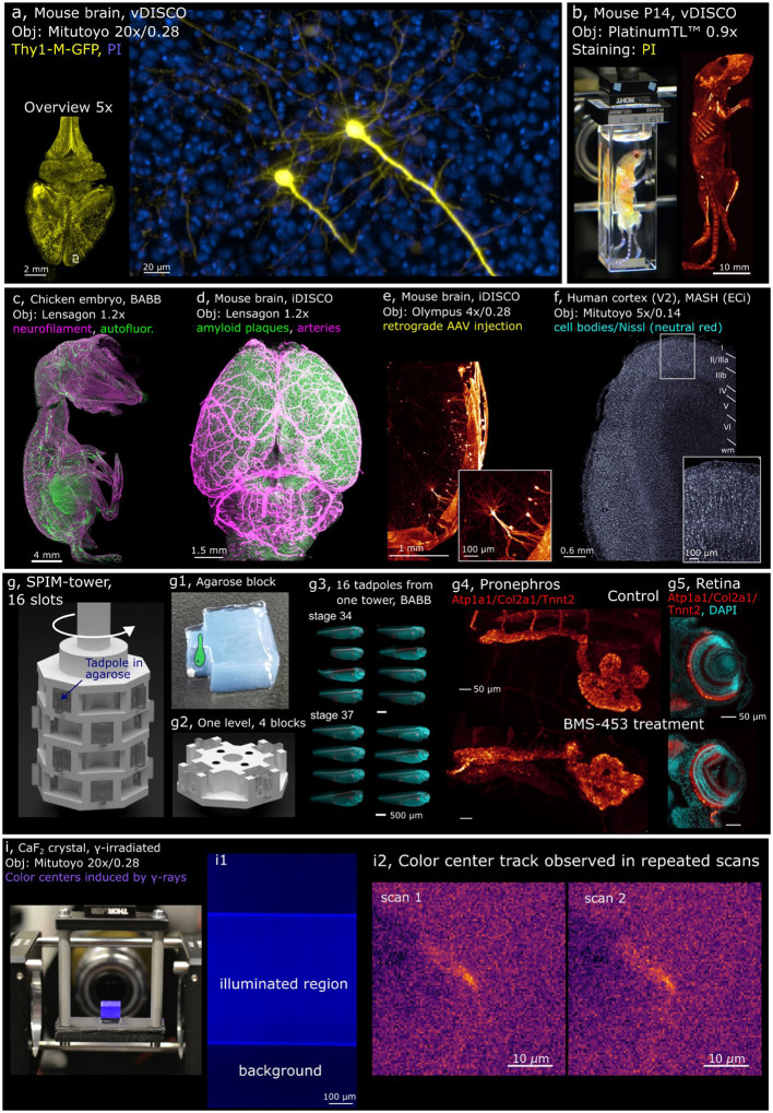
Fig. 3. Examples of Benchtop mesoSPIM applications.
a, Two pyramidal cells in the prefrontal cortex imaged at 20x (Thy1-GFP line M, Atto647N - yellow, propidium iodide - blue, cleared with vDISCO), with axons and basal dendrites resolved. b, Whole mouse body imaging at 0.9x magnification (P14 mouse, stained with propidium iodide PI, cleared with vDISCO). c, Peripheral nervous system of a chicken embryo at E9 imaged at 1.2x (neurofilament staining with mouse anti-RMO270, goat anti-Mouse Cy3; autofluorescence, cleared with BABB). d, Mouse brain at 1.2x (APPPS-1 line, amyloid plaques, arterial vessels, cleared with iDISCO). e, Mouse brain at 4x (Vglut2-Cre line, sparse retrograde AAV injection, iDISCO). f, Human brain tissue at 5x (area V2, stained with neutral red, cleared with MASH). g, CAD model of the SPIM-tower sample holder and the imaging results from high-throughput imaging session. g1, a molded agarose block depicting the sample as a cartoon drawing. g2, CAD model of a single layer of the SPIM-tower sample holder g3, Standardized imaging of 16 X. tropicalis tadpoles (stages 34 and 37) treated with BMS-453 (left) or control (right). Samples were stained with DAPI (cyan) and for Atp1a1/Col2a1/Tnnt2 (red), embedded in individual agarose blocks using the SPIM-mold; g4, pronephros at stage 42 (top: control, bottom: treated), g5 retina (top: control, bottom: treated). BMS-453 treatment affects both kidney and retinal development. i, Irradiated CaF2 crystal imaged at 20x magnification (color centers induced by gamma irradiation at 5 MRad, polished crystal, no clearing); i1, raw image (color-coded blue) showing SPIM-illuminated region vs background fluorescence in the irradiated CaF2 crystal; i2, Candidate track of color centers observed in repeated scans of the irradiated CaF2 crystal. The structure appears across five planes (z-step 3 μm), so the maximum intensity stack is shown for each scan. In all panels, MIPs over multiple planes are shown.

