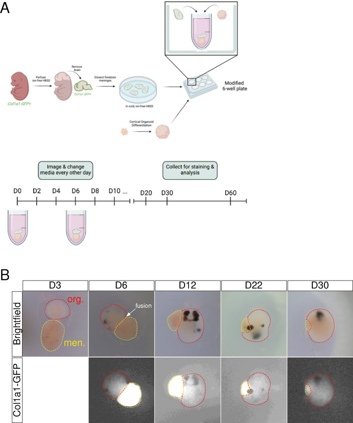
Figure 2: Leptomeningeal-neural organoid (LMNO) fusion culture establishment and growth.
(A) Graphical depiction of leptomeningeal dissection and establishment of LMNO fusions cultures. (B) Representative stereoscope images of LMNO fusions throughout time in culture. Red-dashed lines highlight organoid component, yellow-dashed lines and Col1a1-GFP fluorescence distinguishes meningeal component.

