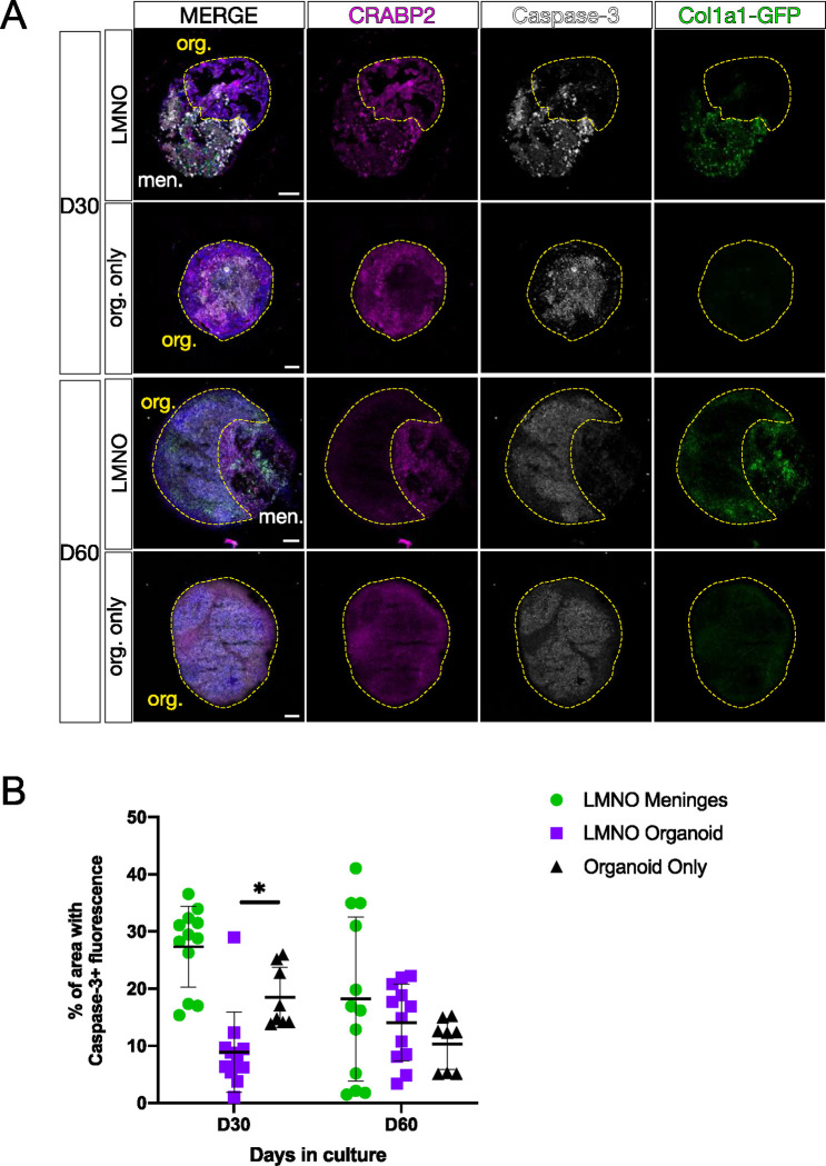
Figure 4: Neural organoids in LMNO fusions show reduced levels of apoptosis.
(A) Images showing Caspase-3 (white) and CRABP2 (magenta) staining in LMNO fusions and non-fused organoids (org. only) at 30 and 60 days in culture, Col1a1-GFP (green) labeling fibroblasts. Yellow-dashed lines outline organoid compartment. Yellow-dashed lines distinguish organoid compartment (org.) from meninges (men.). All scale bars 100μm. (B) Quantification of percentage of area of organoid and meningeal compartments with Caspase-3 fluorescence at 30 and 60 days in culture; meninges in the LMNO fusion (green circles), LMNO fusions (purple squares), non-fused organoid (black triangle). Two-way ANOVA with multiple comparisons revealed significant differences between LMNO fusion versus organoid only at D30 (p = 0.0423) (p< 0.05, *).

