
Figure 5: LMNO fusions recapitulate in vivo Cxcl12-dependent REELIN+ Cajal-Retzius cell migration.
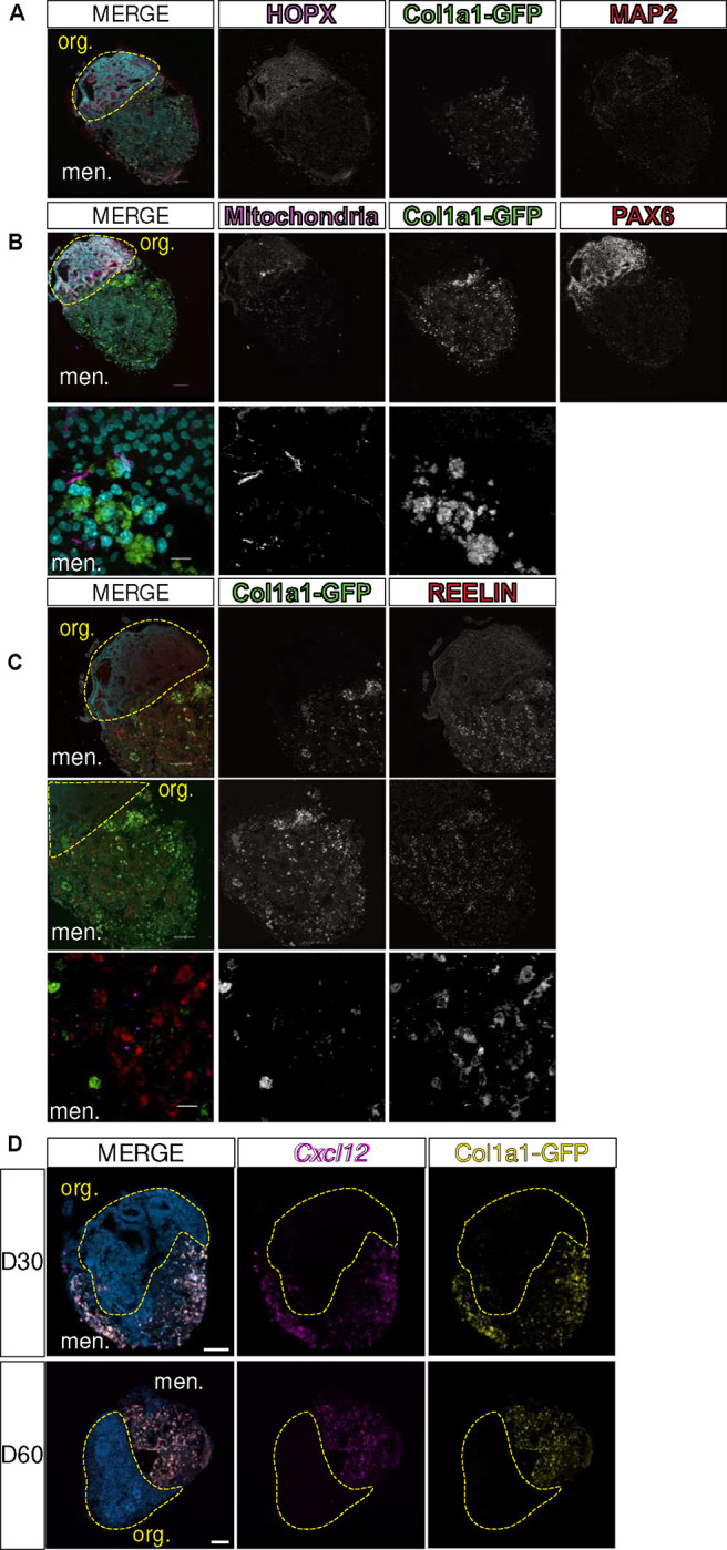
(A-B) Images of LMNO fusions labeled with (A) neuronal markers HOPX (magenta)/MAP2 (red), (B) progenitor marker PAX6 (red) and human mitochondria (magenta). Top image scale bar 100μm and bottom image scale bar 10μm. (C) Cajal-Retzius cell marker REELIN (red); Col1a1-GFP marking meningeal fibroblasts (green). Top 2 images scale bar 100μm and bottom image scale bar 10μm. (D) Images of in situ detection of Cxcl12 (magenta) in LMNO fusions at 30 and 60 days in culture, Col1a1-GFP marking meningeal fibroblasts (yellow). Yellow-dashed lines distinguish organoid compartment (org.) from meninges (men.). All scale bars 100μm.