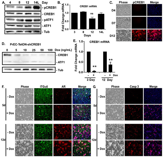
Figure 2: CREB1 is required for luminal cell survival.
Confluent PrECs were induced to differentiate with 2ng/ml KGF plus 5nM R1881 for 3-14 days. ‘L’ indicates only the luminal cells were analyzed. A) Total CREB1, ATF1, and activated levels, i.e., phosphorylation at Ser 133 (pCREB1, pATF1), measured by immunoblotting. Tubulin (Tub) served as loading control. B) Levels of CREB1 mRNA measured by qRT-PCR. C) Differentiated cultures immunostained with anti-pCREB (Ser133) antibody and DNA counterstained with Hoescht (Merge) and imaged by phase or fluorescence microscopy. D,E) Dox concentration required to suppress CREB1 protein and mRNA expression in PrECs expressing Tet-inducible shRNA (PrEC-TetON-shCREB1) assessed by D) immunoblotting at Day 12 and E) qRT-PCR at Day 3 and 12, **p<0.01; n=3; error bars SD. F,G) PrEC-TetON-shCREB1 cells induced to differentiate for 5 or 12 days in the absence (-Dox) or presence (+Dox) of 25ng/ml doxycycline. Cultures immunostained for integrin α6 (ITGα6, basal marker), AR (luminal marker), cleaved caspase 3 (Casp 3) and DNA counterstained with Hoescht (Merge) imaged by phase contrast and fluorescence microscopy.

